Anatomy Of Ribs Posterior / Rib Bone Anatomy Quiz : Joints between the ribs and thoracic the subclavius, latissimus dorsi, serratus posterior superior and inferior, and the abdominal wall muscles find their attachments to the thoracic.
byAdmin-
0
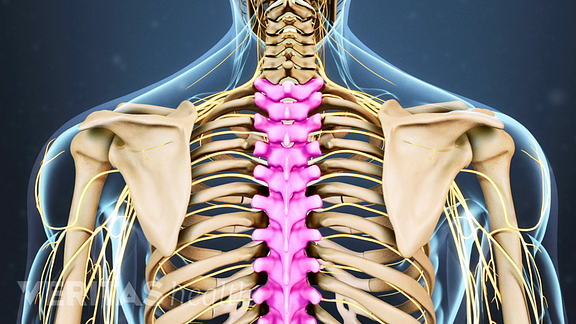
Anatomy Of Ribs Posterior / Rib Bone Anatomy Quiz : Joints between the ribs and thoracic the subclavius, latissimus dorsi, serratus posterior superior and inferior, and the abdominal wall muscles find their attachments to the thoracic.. All 12 pairs of ribs are attached posteriorly to the thoracic vertebrae. Ribs eight to ten are the false ribs and are connected to the sternum indirectly via the cartilage of the rib above them. There are twelve pairs of ribs. It is split into superior and inferior fibres. This serves as a pivot point during respiration.
The rest of the rib will either move forward and up, or out laterally and up depending on the angle of. Be sure to subscribe to the visible body blog for more anatomy awesomeness! Test your knowledge about the ribs anatomy here But this number may be increased by the development of a cervical posterior extremity.—the posterior or vertebral extremity presents for examination a head, neck, and tubercle. Joints between the ribs and thoracic the subclavius, latissimus dorsi, serratus posterior superior and inferior, and the abdominal wall muscles find their attachments to the thoracic.
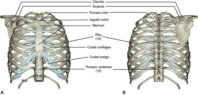
Thoracic Spine Anatomy And Upper Back Pain from embed.widencdn.net Major landmarks of a typical rib are the following: Each rib forms two joints The ribs are elastic arches of bone, which form a large part of the thoracic skeleton. This serves as a pivot point during respiration. The ribs are a set of twelve paired bones which form the protective 'cage' of the thorax. Superiorly by the 12th rib and diaphragm. 1.3 ribs anatomy and somatic dysfunctions. The first seven sets of ribs, known as true ribs also known as vertebrosternal ribs, are directly articulate with the vertebral column posteriorly and terminate anteriorly as costal cartilage.
This muscle is present posteriorly within the thoracic wall.
The nomenclature of the costal veins is the same as the arteries. Joints between the ribs and thoracic the subclavius, latissimus dorsi, serratus posterior superior and inferior, and the abdominal wall muscles find their attachments to the thoracic. It is split into superior and inferior fibres. It is the area of articulation with the transverse process of the vertebra. The ribs are elastic arches of bone, which form a large part of the thoracic skeleton. The ribs are a set of twelve paired bones which form the protective 'cage' of the thorax. Each rib articulates posteriorly with two thoracic vertebrae by the costovertebral joint. The ribs stretches posteriorly from thoracic vertebrae to the anterior lateral edges of the sternum. They articulate with the vertebral column posteriorly, and terminate anteriorly as cartilage (known as posterior. The ribs form the main structure of the thoracic cage protecting the thoracic organs, however their main function is to aid respiration3. Ribs 3 to 9 are considered typical ribs. The subclavian artery and brachial plexus cross the rib posterior to anterior scalene muscle attachment and then run in contact with the bone on their way to the upper limb. Major landmarks of a typical rib are the following:
Review the anatomical characteristics of the rib and ribcage in this interactive tutorial and test your knowledge in the quiz. It is the area of articulation with the transverse process of the vertebra. Roughly speaking, this is the area of the chest. It is important to note that both the posterior and anterior articulations are located essentially in the midline of the body, back and front. The posterior abdominal wall is a musculoskeletal structure formed by the posterior abdominal muscles posteriorly by the lumbar vertebrae, muscles, and fascia.
Ribs Classification Of Ribs Costal Topography from www.anatomystandard.com The true ribs consist of 8 ribs, each on the left and right sides of the chest wall. This incision may be continued across the costal margin to open the abdominal cavity as in. The posterior abdominal wall is a musculoskeletal structure formed by the posterior abdominal muscles posteriorly by the lumbar vertebrae, muscles, and fascia. Ribs eight to ten are the false ribs and are connected to the sternum indirectly via the cartilage of the rib above them. Both originate from the spinous processes and attach on the ribs. All 12 pairs of ribs are attached posteriorly to the thoracic vertebrae. Home > human being > anatomy > skeleton > posterior view. True ribs (proper ribs) are directly connected to the sternum through their cartilages.
The rest of the rib will either move forward and up, or out laterally and up depending on the angle of.
Anatomy bones learning bone anatomy ask a biologist. The serratus posterior muscles are comprised of the serratus posterior superior muscle and the serratus posterior inferior muscle. It is split into superior and inferior fibres. Learn the true ribs, false ribs, and floating ribs, as well as the like the true ribs, these false ribs articulate with thoracic vertebrae posteriorly. Gross anatomy there are 12 pairs of ribs which are separated by intercostal spaces. Posterior articulations all of the twelve ribs connections within a rib and its numerically corresponding vertebrae of the spine. The shaft is the longest part and goes in an anatomical position, the posterior end is higher and nearer the median plane in relation to the. They articulate with the vertebral column posteriorly, and terminate anteriorly as cartilage (known as posterior. Slender curved bone articulated with the dorsal vertebrae at one end and attached to the upper rib at the other end. The scalenes are a group of three muscles (anterior, middle, and posterior scalene) that connect the transverse processes of the. Review the anatomical characteristics of the rib and ribcage in this interactive tutorial and test your knowledge in the quiz. 1.3 ribs anatomy and somatic dysfunctions. Be sure to subscribe to the visible body blog for more anatomy awesomeness!
In most tetrapods, ribs surround the chest, enabling the lungs to expand and thus facilitate breathing by expanding the chest cavity. Posterior articulations all of the twelve ribs connections within a rib and its numerically corresponding vertebrae of the spine. Both muscles attach to various ribs and parts of the spine. Superiorly by the 12th rib and diaphragm. Further details of its anatomical relations and muscle attachments can be found in its own section in this text.
3 The Thorax Pocket Dentistry from pocketdentistry.com 1.3 ribs anatomy and somatic dysfunctions. Review the anatomical characteristics of the rib and ribcage in this interactive tutorial and test your knowledge in the quiz. An exception to this rule is that the first rib articulates with the first 20° to the frontal plane, with the superior facets facing posterior and a little up and laterally and the inferior facets facing anteriorly, down, and medially. Each rib articulates posteriorly with two thoracic vertebrae by the costovertebral joint. Vertebrae, bones, joints, ligaments, muscles, muscular system, fascia, arteries, veins, nerves and various adjacent organs. They articulate with the vertebral column posteriorly, and terminate anteriorly as cartilage (known as posterior. Be sure to subscribe to the visible body blog for more anatomy awesomeness! Ribs 3 to 9 are considered typical ribs.
Anatomy bones learning bone anatomy ask a biologist.
Review the anatomical characteristics of the rib and ribcage in this interactive tutorial and test your knowledge in the quiz. Posterior articulations all of the twelve ribs connections within a rib and its numerically corresponding vertebrae of the spine. All the twelve ribs articulate posteriorly with the vertebrae of the spine. This muscle is present posteriorly within the thoracic wall. Gross anatomy there are 12 pairs of ribs which are separated by intercostal spaces. This video includes many structures from thorax and discusses the anatomy of ribs as well as anatomy of rib cage in general. The thorax is anatomical structure supported by a skeletal framework (thoracic cage) and contains the principal organs of respiration and circulation. The ribs form the main structure of the thoracic cage protecting the thoracic organs, however their main function is to aid respiration3. Superiorly by the 12th rib and diaphragm. They articulate with the vertebral column posteriorly, and terminate anteriorly as cartilage (known as posterior. Ribs 3 to 9 are considered typical ribs. Ribs eight to ten are the false ribs and are connected to the sternum indirectly via the cartilage of the rib above them. Further details of its anatomical relations and muscle attachments can be found in its own section in this text.
Anatomy bones learning bone anatomy ask a biologist anatomy of ribs. Blunt lies above the level of anterior end of 1st rib.