Shoulder Joint Anatomy Diagram Easy / تحلیل و بررسی اوردواهاستاسانا - Iyengar Yoga Shiraz - Due to the tension by the anterior band of the inferior ghl labral teras will be easier to detect.
byAdmin-
0
Shoulder Joint Anatomy Diagram Easy / تحلیل و بررسی اوردواهاستاسانا - Iyengar Yoga Shiraz - Due to the tension by the anterior band of the inferior ghl labral teras will be easier to detect.. Three bones come together at the shoulder joint. The shoulder joint is formed where the humerus (upper arm bone) fits into the scapula. Shoulder joint of human body anatomy infographic diagram with all parts including bones ligaments muscles bursa cavity capsule cartilage membrane for medical science education and health care. Glenohumeral joint commonly called the shoulder joint, the glenohumeral joint helps you move your shoulder forward and backward. Sometimes it is easier to understand anatomy when you look at a visual representation.
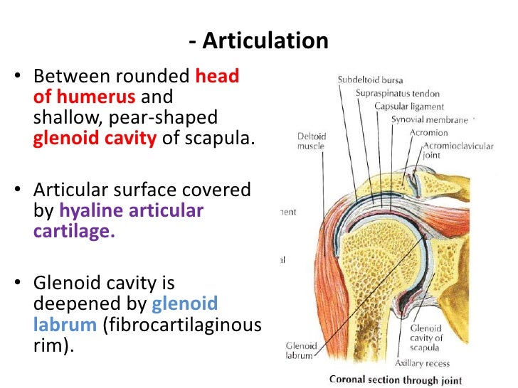
The shoulder is one of the largest and most complex joints in the body. Atlas of the anatomy of the joint of the shoulder on a ct arthrogram in axial, coronal, and sagittal sections, on a 3d images and on conventional athrogram. The glenohumeral, or shoulder, joint is a synovial joint that attaches the upper limb to the axial skeleton. Comprising of numerous ligamentous and muscular structures, the only the joint capsule attaches proximal to the glenoid fossa and attaches further distally to the anatomical neck of the humerus. This attaches to the upper and posterior end of the clavicle and cartilage of the 1st rib purpose:
Shoulder Anatomy Image - Anatomy Drawing Diagram from www.uthscsa.edu Learn more about the shoulder joint anatomy. The glenohumeral joint (shoulder joint) is a synovial ball and socket articulation anatomy ▶ upper limb ▶ joints ▶ shoulder joint (glenohumeral joint). Hey caterina, you somewhat demonstrated the problem of considering the shoulder joint as part of. Start studying shoulder joint anatomy. 8 name the arteries and the nerves that supply shoulder joint. Shoulder joint of human body anatomy infographic diagram with all parts including bones ligaments muscles bursa cavity capsule cartilage membrane for medical science education and health care. Chevy impala radio wiring diagram. Equally extensive are the muscles affecting the shoulder movement, including:
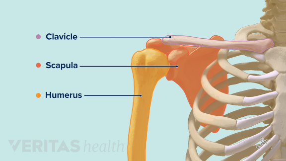
These two joints work together at the arm to allow the shoulder to move in a large circle and to rotate around its axis.
Hey caterina, you somewhat demonstrated the problem of considering the shoulder joint as part of. Atlas of the anatomy of the joint of the shoulder on a ct arthrogram in axial, coronal, and sagittal sections, on a 3d images and on conventional athrogram. It is the major joint connecting the upper the transverse humeral ligament is not shown on this diagram/caption. It has the largest range of motion out of all the joints in the body and consists of three bones. This diagram here just shows the joint capsule itself. Simply put, the shoulder, or shoulder joint, is the connection of the upper arm and the thorax. • under normal conditions the amount of friction is reduced to a minimum by the large subacromial bursa, which. Home > blog > anatomy > shoulder anatomy: Comprising of numerous ligamentous and muscular structures, the only the joint capsule attaches proximal to the glenoid fossa and attaches further distally to the anatomical neck of the humerus. Click now and learn everything about its anatomy and function at kenhub! All about the shoulder muscles. These two joints work together at the arm to allow the shoulder to move in a large circle and to rotate around its axis. The shoulder is one of the largest and most complex joints in the body.
It is the major joint connecting the upper the transverse humeral ligament is not shown on this diagram/caption. The shoulder joint is the connection between the chest and the upper extremity. • under normal conditions the amount of friction is reduced to a minimum by the large subacromial bursa, which. This diagram here just shows the joint capsule itself. Shoulder joint of human body anatomy infographic diagram with all parts including bones ligaments muscles bursa cavity capsule cartilage membrane for medical science education and health care.
تحلیل و بررسی اوردواهاستاسانا - Iyengar Yoga Shiraz from iyengaryogashiraz.com Due to the tension by the anterior band of the inferior ghl labral teras will be easier to detect. 1 this mobility provides the upper extremity with tremendous range of motion such as adduction, abduction, flexion, extension, internal rotation, external rotation, and 360° circumduction in. These two joints work together at the arm to allow the shoulder to move in a large circle and to rotate around its axis. See more ideas about joints anatomy, shoulder joint, shoulder joint anatomy. Looking for quizzes, videos, articles and an. Describe the structure of the shoulder should begin with bone parts that include: Three bones come together at the shoulder joint. Diagram of the human shoulder joint, back view.
This diagram here just shows the joint capsule itself.
Shoulder joint of human body anatomy infographic diagram with all parts including bones ligaments muscles bursa cavity capsule cartilage membrane for medical science education and health care. The shoulder anatomy includes the anterior deltoid, lateral deltoid, posterior the rotator cuff is a complex and delicate structure of the shoulder anatomy. The shoulder is one of the largest and most complex joints in the body. This diagram here just shows the joint capsule itself. Atlas of the anatomy of the joint of the shoulder on a ct arthrogram in axial, coronal, and sagittal sections, on a 3d images and on conventional athrogram. As a ball and socket synovial joint, there is a wide range of. You can see it enclosing the glenohumeral joint and you can see its attachment on the anatomical neck that's the shoulder joint. Describe the structure of the shoulder should begin with bone parts that include: The left shoulder and acromioclavicular joints, and the proper ligaments of the scapula. Speed drawing shoulder ligaments подробнее. Diagram of the human shoulder joint, back view. Robin smithuis and henk jan van der woude. Normal anatomy, variants and checklist.
Related online courses on physioplus. The shoulder joint is vulnerable to dislocations from sudden jerks of the arm, especially in children before strong muscles have developed. You can see it enclosing the glenohumeral joint and you can see its attachment on the anatomical neck that's the shoulder joint. Glenohumeral joint commonly called the shoulder joint, the glenohumeral joint helps you move your shoulder forward and backward. Posted on december 13, 2018december 12, 2018.
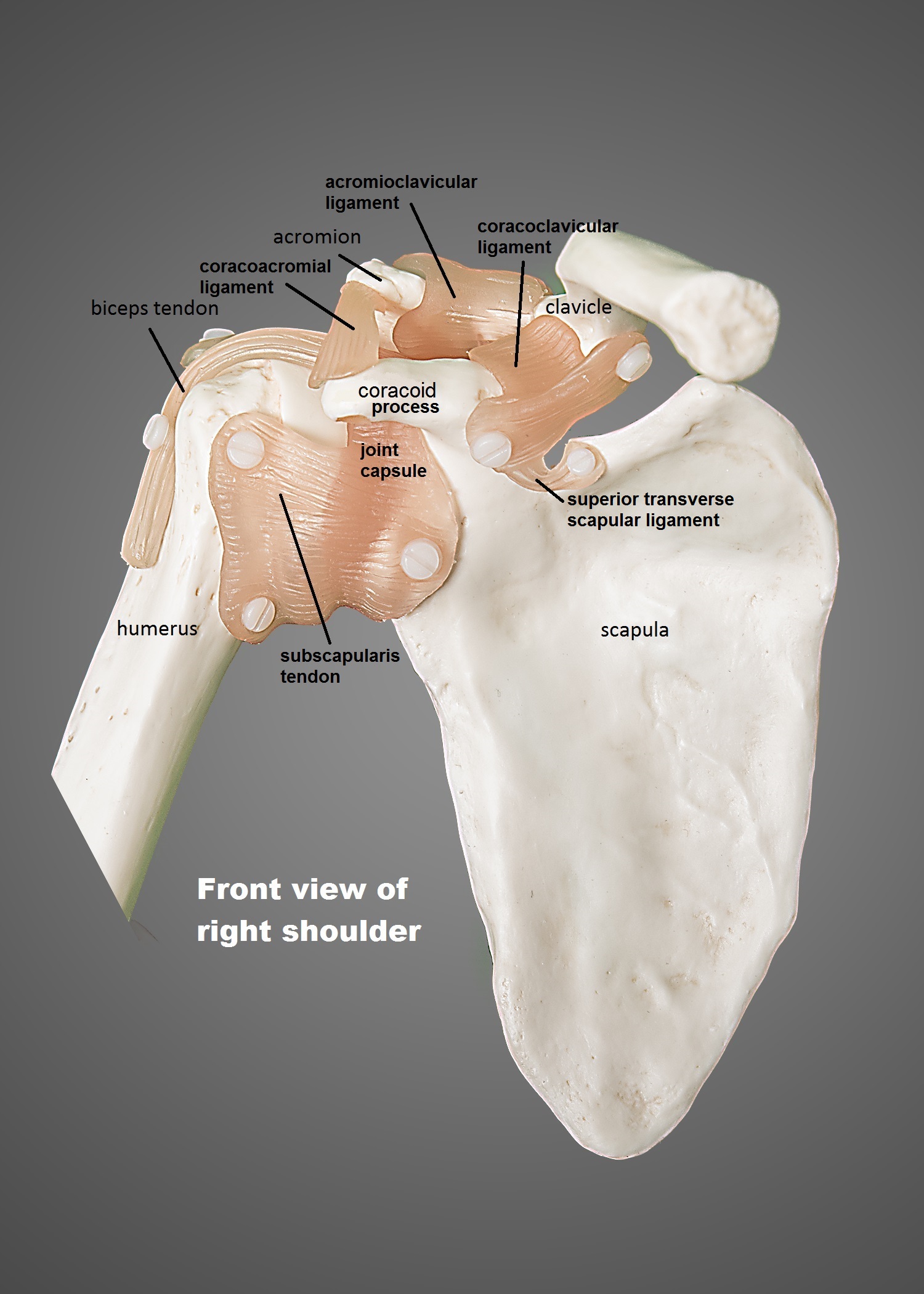
Anatomy Of The Glenohumeral Joint - Anatomy Drawing Diagram from image.slidesharecdn.com License image the shoulder joint ligaments shown are the acromioclavicular ligament, coracoacromial ligament, coracohumeral ligament, coracoclavicular ligament, and the articular capsule or glenohumeral. This attaches to the upper and posterior end of the clavicle and cartilage of the 1st rib purpose: Glenohumeral joint commonly called the shoulder joint, the glenohumeral joint helps you move your shoulder forward and backward. In common usage, shoulder joint mostly refers to the glenohumeral joint, the major joint of the shoulder but can also include acromioclavicular joint. Learn vocabulary, terms and more with flashcards, games and other study tools. The students must thoroughly study the shoulder joint as it usually undergoes recurrent dislocations and is the most common joint to dislocate. Posted on december 13, 2018december 12, 2018. Speed drawing shoulder ligaments подробнее.
Describe the structure of the shoulder should begin with bone parts that include:
Click now and learn everything about its anatomy and function at kenhub! Hey caterina, you somewhat demonstrated the problem of considering the shoulder joint as part of. Posted on december 13, 2018december 12, 2018. The shoulder is one of the largest and most complex joints in the body. See more ideas about joints anatomy, shoulder joint, shoulder joint anatomy. Shoulder joint of human body anatomy infographic diagram with all parts including bones ligaments muscles bursa cavity capsule cartilage membrane for medical science education and health care. Atlas of the anatomy of the joint of the shoulder on a ct arthrogram in axial, coronal, and sagittal sections, on a 3d images and on conventional athrogram. The human shoulder is the most mobile joint in the body. Three bones come together at the shoulder joint. The glenohumeral joint (shoulder joint) is a synovial ball and socket articulation anatomy ▶ upper limb ▶ joints ▶ shoulder joint (glenohumeral joint). Diagram of the human shoulder joint, back view. Home > blog > anatomy > shoulder anatomy: Describe the structure of the shoulder should begin with bone parts that include:
The glenohumearal joint has a greater range of motion than any other joint in the body shoulder anatomy diagram. Related online courses on physioplus.